Dr Joepaul Joy, Dr Sheela Nampoothiri,Dr Dhanya Yasodharan(Paediatric Genetics), Dr C Jayakumar
Twenty days old male ba from Kanyakumari, first child of nonconsanguenous parentage delivered LSCS due to breech with birth weight- 3.15kg
Mother was on treatment for hypothyroidism she had oligohydramnios
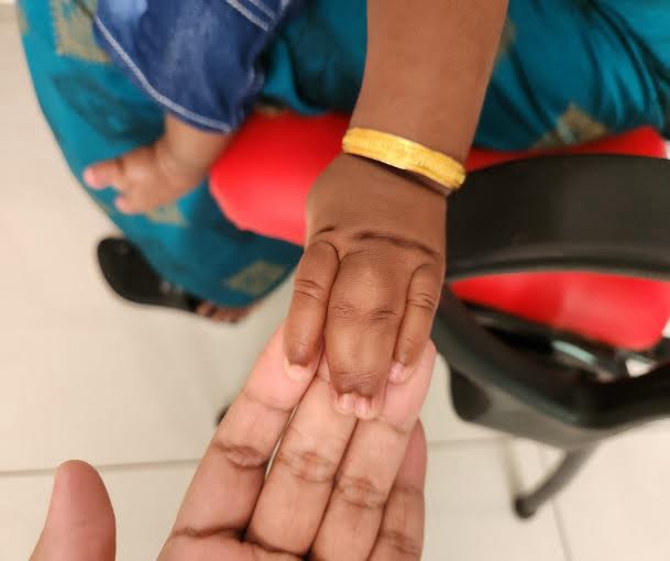
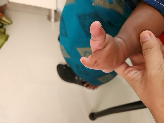
Postnatally ba was observed to have syndactyly of bilateral 3rd and 4th fingers, bilateral ectrodactyly of feet. Ba was admitted on day 7 of life in view of poor feeding and weight loss.
Echo and USG Abdomen done were normal. He was discharged after 2 days on breastfeed and topfeed. In view of anatomical anomaly of hands and feet ba was brought to AIMS for orthopaedic consultation.
On examination ba was active,alert with stable vitals.Physical exam of the ba was other wise normal
Ba had weight of 2.9kg(<-3SD), length of 53cm(b/w 0 and -2 SD) and HC-37cm(b/w 0 and -1 SD).Head to foot examination revealed low set posterior rotated pinna, bilateral syndactyly of 3rd and 4th fingers, bilateral ectrdactyly of feet(left foot has 2 toes and right foot has 3 toes with syndactyly of 4th and 5th toes. Systemic examination werewithin normal limits. As per Plastic surgery opine Syndactyly release and cleft foot repair after 1 year of agewas planned
Genetics dept advised to do Microarray which was normal following which WES was done which showed homozygous mutation in WNT10B gene leading to split hand/foot malformation.
Parents are heterozygous carriers. Ba is advised to review after one year of age.


Split hand/foot malformation is a limb abnormality that is present at birth. It is characterized absence of certain fingers and toes (ectrodactyly) that suggests a claw like appearance and webbing of fingers and toes may also be present. SHFM can be inherited as a single abnormality or as part of a syndrome that includes other characteristics. Most people with SHFM have fewer than five fingers or toes on a hand or foot (oligodactyly). A smaller proportion of individuals have finger fusing (syndactyly) of multiple fingers on the hand.
This is often referred to as the “lobster claw” variety. SHFM can be inherited in autosomal dominant pattern, autosomal recessive pattern and also x-linked pattern. It can also occur as a result of a random (sporadic) mutation during fertilization or embryonic development. It is usually diagnosed physical features present at birth. Genetic testing for genes can be done to confirm the diagnosis. Reconstructive surgery can be performed to improve function and appearance when applicable. Prosthetics are also available.
Take home message: All babies with limb anomalies at birthshould be extensively evaluated for better management and outcome.
