Dr.Subbulakshmi.P.S*, Dr. Jayakumar.C*, Dr.Praveena Bhaskaran*, Dr.Suhaas Udaykumar**
*Department of Paediatrics, **Department of neurosurgery
AIMS, Kochi
Two years old female child, 1st child of non-consanguineous marriage, apparently normal 3 weeks back, developed pain in the neck region which was insidious in onset and radiating to bilateral shoulder region after which mother noticed a small insignificant swelling at the back of neck which was managed conservatively. After 5 days, the child developed inability to turn her neck to the right side for which she was taken to a near hospital which was again managed symptomatically. The child did not show any improvement and the symptoms persisted and the mother also noticed weakness in the right upper and lower limb. The child became immobile with persistent torticollis. Birth and past history was uneventful. No relevant family history was also noted. Immunized till age and age appropriate milestones were attained.
No history of fever/ loose stools/ vomiting/ recent URI/ seizures/ altered sensorium/ normal bladder and bowel habits. No H/O trauma noted.
On examination, the child was not sick-looking and oriented to time, place and person. She was vocalising but can neither turn her head nor move her right upper and lower limbs. GCS was 15 and pupils were reactive. Bulk was normal and power was decreased in the right UL and LL (1/5) and normal on the left. Deep tendon reflexes were very brisk on the right limbs and normal on the left. Plantars were down-going and the child had a hypotonia over the right limbs. No cerebellar signs or meningeal signs noted. No stigmata of phakomatoses. Other systemic examinations were within normal limits.
DIFFERENTIAL DIAGNOSIS:
1. Paediatric stroke
2. Space occupying lesions
3. Guillian barre syndrome
4. Traumatic myelitis
5. Poisoning
6. Retropharyngeal abscess
INVESTIGATIONS:
Hb: 12 g/dl
PCV: 36 %
Platelets: 327 ku/ml
TC: 9.25 ku/ml
DC: 35 N/55 L/1.9 E
CRP : 37.85 mg/L
PT/INR : 16.2/14.7/1.11 sec
APTT: 28.1/30.5 s
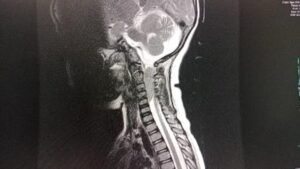
MRI Spine: Solid lobulated intradural extramedullary tumour in the cervical region, located anterolateral and to the right of the cervical cord, compressing the cord- C2-C5 level, 36 X 16 X 12mm. Lesion is likely to represent a dural based tumour like a meningioma. No extraspinal extension. No other similar lesions in the cerebral parenchyma or in the rest of the spinal canal.
The child under-went a Laminoplasty C2- C6 and near total excision with Intraoperative neuromonitoring under GA immediately after MRI report and post-operatively she went into a septic shock which was treated as per protocol. After stabilization, a post-op MRI spine was taken which showed no residual lesion at the post operative site with mildly effaced posterior thecal space and no cord compression. Biopsy of the resected tumor mass showed C2-C5 IDEM, poorly diffrentiated high grade Malignant Neoplasm with negative INI1 in view of which a possibility of Atypical Teratoid/Rhabdoid tumour is considered and hence planned to start radiotherapy followed chemotherapy. The child is discharged with stable vitals and kept under close follow-up.

MRI head and neck image before surgery showing a SOL in C2-C5 site.

MRI spine image after surgery showing clearing.
DISCUSSION: Acute onset hemiplegia in a 3-year-old is a serious condition that requires urgent medical attention.
Possible Causes:
1. Stroke
– Arterial or venous malformations
– Clotting disorders
– Congenital heart defects
– Infections
2. Infections:
– Meningitis
– Encephalitis
3. Trauma
4. Brain Tumors
5. Seizures
6. Neurovascular Events : Conditions such as moya moya disease, which causes progressive stenosis of the brain’s blood vessels, can lead to stroke-like symptoms.
Evaluation and Diagnosis
– Medical History and Physical Examination: Initial assessment to understand the onset, associated symptoms, and any potential triggers or underlying conditions.
– Neuroimaging: MRI or CT scans of the brain to identify abnormalities such as stroke, tumors, or hemorrhage.
– Lumbar Puncture: If infection is suspected, cerebrospinal fluid analysis may be necessary.
– Blood Tests: To check for infections, metabolic or clotting disorders.
Treatment
-Acute Management :
Depends on the underlying cause but may include medication to manage seizures, anticoagulants for stroke, or antibiotics for infections.
– Supportive Care:
May involve physical therapy, occupational therapy, and speech therapy depending on the severity of the hemiplegia.
– Surgical Intervention : In cases of trauma or tumors, surgical options might be considered.
TAKEHOME MESSAGE:
The outcome depends on the cause, the extent of brain injury, and the timeliness of treatment. Early intervention generally improves the prognosis and can reduce the risk of long-term complications. Immediate medical evaluation is essential to determine the cause and initiate appropriate treatment to optimize outcomes for the child.
