Dr.venumadhav, Dr.Vinayan,DrVyshak (paed Neuro) ,DrC Jayakumar
Amrita Institute of Medical sciences, Kochi
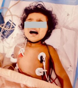
A 1-yr 3-month-old male child presented with high-grade intermittent fever and irritability lasting for 9 days. No pointer symptoms suggestive other system involvement
ITreated at periphery hospital for a urinary tract infection
Due to persistent symptoms, he was referred to AIMS
The child is developmentally normal and immunized for age.
Examination and Investigations:
On examination, the child was febrile and irritable, with neck stiffness. PICCLE normal . Systemic examination was otherwise unremarkable.
.
Differential Diagnosis:
Viral Meningitis/ Encephalitis
Eosinophilic Meningitis
Partially treated Bacterial Meningitis
Viral fever
Partially treated UTI
Autoimmune Encephalitis
Cerebral Vasculitis
Sub Arachnoid haemorrhage
Initial investigations revealed peripheral eosinophilia (14%) with negative CRP, and serological tests for leptospirosis, dengue, brucella, and tropical fever were all negative.
The respiratory viral panel (RVP) was positive for Influenza B, so Oseltamivir was started.
Lumbar puncture was done, which revealed mixed pleocytosis with predominant eosinophilia, elevated protein, and low glucose (Glu-24, protein 104, Eosionophills-50%).
MRI brain imaging showed extensive cortical and subcortical areas of diffuse restriction, focal T2 FLAIR hyperintensities, and leptomeningeal enhancement along the bilateral cerebellar foliae.
Given these atypical MRI findings, CNS vasculitis was suspected, and IVIG was administered. Cultures of CSF and urine showed no growth, and tests for tuberculosis and the meningoencephalitis panel were negative.
The parents reported the presence of snails in the vicinity of their house, leading to the consideration of eosinophilia meningitis as a diagnosis, given the epidemiology, clinical presentation, and laboratory and Imaging findings (Peripheral eosinophilia+ CSF cytology- Eosinophillia, Increased CSF protein with low glucose)
MRI showing
Extensive cortical and Subcortical areas of diffuse restriction


Leptomeningeal enhancement along bilateral cerebellum foliae
TREAMENT
Main stay of treatment is steroids
Initial Management: The child was started on pulse IV Methylprednisolone for 3 days, followed oral steroids (1 mg/kg/day) for 2 weeks duration
Anthelmintic Therapy: Later, Albendazole was administered at 15 mg/kg/day under steroid cover for a total of 2 weeks.
Starting anthelmintic therapy early may cause the worms to die, leading to increased inflammation in the central nervous system
DISCUSSION:
Human infection with Angiostrongylus cantonensis primarily affects the Central Nervous System (CNS), where the larvae migrate after ingestion. In humans, the worms usually die in the CNS, provoking an inflammatory response that contributes to various symptoms. This spectrum of disease, collectively termed Eosinophilic Meningitis (AEM), includes meningitis, encephalitis, encephalomyelitis, and radiculitis, reflecting the different clinical manifestations
1. Clinical Presentation:
o Uncomplicated Meningitis: Most commonly, patients present with symptoms such as severe headache, neck stiffness, photophobia, visual disturbances, hyperesthesia, fatigue, and vomiting. These symptoms typically manifest within 1 to 4 weeks following the ingestion of contaminated food.
Severe Neurological Symptoms: In cases of heavy infestation, patients may develop encephalitis, radiculomyelitis, or even coma and death.
o Ocular Symptoms: Some patients primarily present with unilateral visual disturbances, with minimal systemic symptoms.
2. CSF and Blood Findings:
o CSF Eosinophilia: The hallmark of AEM is eosinophilia in the CSF, which may not be present at the first lumbar puncture but often develops later in the course of the disease.
o Peripheral Blood Eosinophilia: This may also be observed, typically peaking about two weeks after symptom onset.
o MRI: Imaging is often nonspecific but can reveal leptomeningeal enhancement, brain swelling, or subcortical white matter changes.
o Molecular Testing: PCR assays for A. cantonensisDNA hold promise for early and accurate diagnosis, though they are not widely available.
3. Exposure History:
o Food and Travel History: A thorough history of ingestion of raw or undercooked intermediate or paratenic hosts, such as snails, slugs, or contaminated produce, is critical..
Treatment
The treatment of AEM is not well-established and remains controversial due to the variability in disease severity and the complexity of its pathophysiology.
1. Supportive Care:
o Serial Lumbar Punctures: Frequent lumbar punctures can alleviate symptoms reducing intracranial pressure, particularly in cases of severe headache.
2. Corticosteroids:
o Prednisolone: High-dose corticosteroids have been shown to reduce symptom duration and severity in milder cases of AEM.
3. Anthelmintics:
o Albendazole: This is the most studied anthelmintic, with some evidence suggesting it reduces the duration of symptoms, particularly when combined with corticosteroids.
