
Dr.Terencia/Dr.Neeraj/ Dr. Manoj/ Dr.Rema/ Dr. C Jayakumar.
Amrita Institute Of Medical Sciences, Kochi.
A 7 year old female child was referred from outside hospital with chief complaints of high grade intermittent fever spikes associated with facial puffiness and periorbital swelling of 1 week duration. . URE showed proteinuria and USG Abdomen showed bulky kidneys with marginally increased echotexture and Left sided pleural effusion. Child was initially managed symptomatically in terms of nephrotic syndrome in an outside hospital. CT taken for further evaluation showed enlarged thymus with gross left pleural effusion with passive collapse of left lung. Child was managed with steroids and supportive care but since symptoms worsened and child further developed breathing difficulty she was referred to AIMS for further management
On examination child was sick looking, febrile and tachypneic with no desaturation. She had reduced air entry(L>R) with dull note on percussion and scattered b/l crepitations. She was started on oxygen support, IV antibiotics and other supportive medication. Chest x ray showed mediastinal mass with pleural effusion. Routine investigations showed Hb- 10.2g/dl, PLT – 626 ku/ml , NEU -76.7% , WBC -10.50 ku/ml , creat- 1.15 , urea-60.8 , phos- 6.7 , uric acid-13.5 .
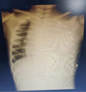
Figure-1-CXR-Mediastinal mass with left side massive pleural effusion
DIFFERENTIAL DIAGNOSIS-
1.Burkitt Lymphoma
2.Acute Myeloid Leukemia
3.Acute undifferentiated Leukemia
4.Chronic Myeloid leukemia
5.Infectious diseases and other causes of lymphocytosis and/or aberrant lymphocytes
Patient was advised hyperhydrarion in view of the tumor lysis and was started on allopurinol and rasburicase for the same. Pediatric cardiology consultation was done to rule out any pericardial effusion as mediastinum shift to right was visible on CXR. ECHO showed left sided massive pleural effusion and good biven-tricular function and no pericardial effusion. In view of the persistent tachypnea the pleural fluid was drained therapeutically and was sent for evaluation.

Figure 2-CXR after pleural fluid drainage
Pleural fluid flow cytometry had no leukemic cells . Pleural fluid cytology had shown lymphocyte cell rich effusion .Gram stain, culture and gene x pert of the pleural fluid was negative. Lymph node biopsy of supraclavicular lymph node was done which had shown lymphoid infiltrate . IHC of the lymph node biopsy had shown positive for CD3,Tdt and negative for CD20 with Ki 67 index-95% cells which are double positive for CD4 and CD8 and suggestive of T cell lymphoblastic lymphoma. Patient parents were explained regarding the disease and the prognosis and chemo-therapy consent was taken .
Patient was started on the oral prednisolone and was given 3 doses of injection cyclophosphamide totally. During the course of hospitalization since she developed further fever spikes and Blood culture was positive for Acinetobacter, IV antibiotics were hiked up according to the sensitivity index. Bone marrow aspiration done and showed particulate cellular marrow with trilineage maturation. Karyotype: 46,XX. Bone Marrow Biopsy showing Hypocellular marrow with trilineage maturation. PICC line insertion was done with groshong catheter. LP with CSF cytology and CSF panel with IT methotrexate was done and showed no atypical lymphoid cells . Chemotherapy vincristine and daunorubicin was started and parents were explained regarding the treatment protocol and consent was obtained
Acute lymphoblastic leukemia/lymphoma (ALL/LBL) refers to hematologic malignancies of lymphoid precursor cells. These entities are described as ALL/LBL because in this setting, leukemia and lymphoma are overlapping clinical presentations of the same disease; the systems for diagnosis and classification do not distinguish between leukemia and lymphoma. Broadly, ALL/LBL is divided into tumors of B cell and T cell lineage; rare tumors of natural killer (NK) cell lineage are also recognized. Immunophenotyping is required to determine the lineage of ALL/LBL because the different subtypes are morphologically indistinguishable.
Most cases of ALL/LBL have cytogenetic and/or molecular abnormalities that are associated with distinctive phenotypes, prognostic features, and/or influence the choice of treatment. The World Health Organization (WHO) classification system uses immunophenotype and cytogenetic/molecular features to define specific categories of ALL/LBL. This topic will review the WHO classification of ALL/LBL and the associated cytogenetic and molecular abnormalities.
Patients are usually males in their teens to early twenties who present with lymphadenopathy in cervical, supraclavicular, and axillary regions (50 percent), or with a mediastinal mass (50 to 75 percent). In most patients the mediastinal mass is anterior, bulky, and associated with pleural effusions. These masses can produce such complications as superior vena cava syndrome, tracheal obstruction, and pericardial effusions (with or without tamponade). The disease tempo is variable with some patients presenting with symptoms progressing slowly over weeks to months and others more acutely.
Less commonly, patients present with extranodal disease (eg, skin, testicular, or bony involvement). Abdominal involvement is very unusual; when it does occur it is primarily in the liver and spleen.
More than 80 percent of patients present with stage III or stage IV disease and almost 50 percent have B symptoms; in the majority, serum lactate dehydrogenase (LDH) levels are elevated. Although the bone marrow is frequently uninvolved at presentation, approximately 60 percent of patients develop bone marrow infiltration and a subsequent leukemic phase indistinguishable from T cell ALL.
Evaluation of the spinal fluid is essential to rule out central nervous system (CNS) involvement, which is frequently seen. Patients with bone marrow involvement have a particularly high incidence of CNS infiltration, which typically takes the form of leptomeningeal disease. Parenchymal involvement of the brain may occur in both T-ALL and B-ALL, but is much less common.
Precursor T-lymphoblastic leukemia/lymphoma (precursor T-LBL), also called precursor T cell acute lymphoblastic leukemia (precursor T cell ALL) is postulated to arise from precursor T lymphoblasts at varying stages of differentiation. The term precursor T cell acute lymphoblastic leukemia (T-ALL) is used if there are >20 percent bone marrow blasts, with or without a mass lesion. For cases with a mass lesion and less than 20 percent bone marrow involvement, the term precursor T cell lymphoblastic lymphoma (T-LBL) is used.
Precursor T cell LBL/ALL occurs most frequently in late childhood, adolescence, and young adulthood, with a male predominance; it comprises 15 percent of childhood and 25 percent of adult ALL and 2 percent of adult non-Hodgkin lymphoma. Peripheral blood smear, bone marrow biopsy, and tissue biopsy can demonstrate lymphoblasts with varying morphology. Precursor T-LBL/ALL is morphologically indistinguishable from precursor B-ALL/LBL.
Key diagnostic studies include histochemical stains, immunocytochemistry, and flow cytometry. These are required to differentiate among precursor T-ALL/LBL, precursor B-ALL/LBL, and other acute leukemias. A number of genetic abnormalities have been identified in patients with precursor T-ALL/LBL; at present, the most prognostically important of these are TP53 mutations.
The diagnosis of precursor T-lymphoblastic leukemia/lymphoma is made based upon the results of a bone marrow biopsy with or without biopsy of other involved tissues, such as the mediastinum. While not yet routine, all acute leukemias seen at our institution are subjected to analysis using a targeted next-generation sequencing platform that covers certain genes that are prognostically important in T-ALL/LBL, such as TP53.
TAKE HOME MESSAGE-Differential Diagnosis of ALL is broad because the clinical presentation is often non specific and lymphoblast morphology is non diagnostic. So its essential to rule out ALL in children with unexplained cytopenias, lymphadenopathy, circulating aberrant lymphoid cells/blasts include viral infection, aplastic anemia other caners and other hematological malignancies include Burkitt lymphoma and acute AML.