
Dr. Terencia, Dr. Madhumitha(Dept of E.N.T), Dr. C Jayakumar
AIMS, KOCHI.
Four year old female child from Russia,tracheostomy depended ,presented with congenital subglottic stenosis since birth and unable to vocalise after a failed laser excision .She has no difficulty in feeding or breathing difficulty.
Child had developed respiratory distress immediately after birth and on further evaluation was diagnosed to have congenital subglottic stenosis and was kept on tracheostomy support since then.
On examination child was alert with surgical site appearing healthy with no swelling, no crepitus. No signs of tube blockage. Systemic examination was within normal limits.
MDCT Neck done and showed focal narrowing with luminal obliteration of subglottic larynx noted for a length of 7mm with elliptical shaped cricoid cartilage. No adjacent soft tissue swelling noted. Right laryngeal ventricle and right pyriform sinus were mildly prominent. Supraglottis and pharynx werenormal. Right upper lobar bronchus directly arising from trachea just above carina. Scanned lung parenchyma normal.

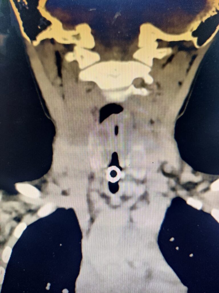
Figure 1 and 2 -MDCT showing Subglottic stenosis
Flexible Fibre Optic Laryngoscopy Under GA was done and showed Grade 4 Subglottic Stenosis and right vocal cord palsy. No stenosis noted in the supraglottic and glottic area. Immediate subglottis showing lateral narrowing leading to complete closure of subglottis and Scope could not be passed beyond. Left vocal cord mobility was present. Tracheostomy tube was removed. Peristomal granulations and Suprastomal granulation were present. Scope could not be passed to visualise supra and infra stomal areas. Tracheostomy tube changed to Bivona cuffless tube. Child was shifted to PICU in stable condition.
She underwent Cricotracheal resection Pediatric Laryngotracheal Surgery Under GA and was kept on ventilator support (nasally intubated) in PICU under sedation. Drain was removed on POD-3. She underwent airway evaluation via Flexible Fibre Optic Laryngoscopy Under GA which showed well healing anastomatic site. She developed perisutural site swelling and crepitus on which was decompressed through drain incision. Bronchoscopy was done as prior to extubation which showed dehiscence in anterior tracheal wall following which Nasotracheal cuffless tube was changed into cuffed tube. She further developed high grade fever with elevated markers. Blood culture showed gram negative bacilli and was started on higher end antibiotics. After stabilisation she was taken up for Relook surgery under GA on and was shifted to PICU where she was intubated and sedated.
Figure 3,4,5 and 6- Cricotracheal
resection Pediatric Laryngotracheal Surgery.
Bronchoscopy was done on POD 3 and anastomotic sites were noticed to be holding with subglottic narrowing immediately inferior to the glottis. She was hence taken up for Flexible Fibre Optic Laryngoscopy Under GA and Kenocort injection was given at the site of narrowing. Child was slowly weaned off ventilation and was extubated to CPAP and was found to be comfortable. Bronchoscopy was repeated and anastomotic site was healing well. She started oral feeds and was tolerating well. As she developed post swallow cough on taking liquids, after Dysphagia consultation started on NG liquid feeds and partially oral for semisolid diet. She was switched to intermittent BiPAP at night and was kept under observation.
Parents were trained with intermittent BiPAP support and was discharged on the same with stable vitals.
CONGENITAL SUBGLOTTIC STENOSIS is a narrowing of the lumen of the cricoid region, possibly caused incomplete canalization of the cricoid ring (diameter less than 4 mm in the term infant and 3 mm in the preterm infant). It presents most commonly as recurrent croup but may cause biphasic stridor in a newborn if severe. It is differentiated from acquired subglottic stenosis the absence of history of trauma or instrumentation and less severe symptoms.Congenital subglottic stenosis typically improves as the larynx grows. Fewer than one-half of the children with this disorder require tracheostomy. Depending upon the severity of the obstruction and the need for supplemental oxygen therapy, congenital subglottic stenosis also may be treated endoscopic division of cartilage with balloon dilation, anterior-cricoid split with posterior cartilage graft, or partial cricotracheal resection
The Bogdasarian and Olson Classification is a classification scheme for posterior glottic stenosis.1
Type 1 → Interarytenoid scar which does not extend to the posterior commissure (between the arytenoids only).
Type 2 → Interarytenoid scar extending to the posterior commissure.
Type 3 → Posterior commissure scar involving one cricoarytenoid joint.
Type 4 → Posterior commissure scar involving both cricoarytenoid joints.
TAKE OVER MESSAGE- Cricotracheal resection (CTR) is a surgery in which the narrowed part of the airway just below the voice box (larynx) is removed and the voice box and windpipe (trachea) are sewn back together. It is also used to treat other airway problems. Cricotracheal resection is performed when cricotracheal stenosis is unresponsive to conservative management, such as balloon dilation and scar excision