Dr. Venumadhav, Dr.suma Balan (PaedRheumatology) Dr. C. Jayakumar
Department of Pediatrics, Amrita Institute of Medical Sciences
Introduction
A sixteen-year-old male presented to the outpatient department with complaints of recurrent episodes of pain and swelling of multiple large joints over the past two years, recurrent painful oral and genital ulcers, and intermittent abdominal pain over the past one year. The oral ulcers healed without scarring. The patient had been treated with multiple alternative and traditional medicines and was currently on homeopathy. There was no history of eye pain, redness, photophobia, fever, cough, headache, vomiting, altered sensorium, or seizures
As swelling and joint pain increased, the child was brought to AIMS for further evaluation and management. He is the second child of a non-consanguineous marriage, developmentally normal with good scholastic performance, and immunized according to the NIS schedule. He had significant failure to thrive with weight below the 3rd percentile (36 kg) and height at the 10th percentile (158 cm).
Examination and Investigations:
On examination, the child appeared tired and uninterested in walking. He was pale with small, multiple, non-tender cervical lymph nodes.
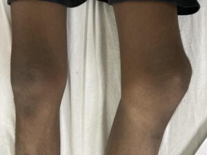
A head-to-toe examination revealed acneiform lesions over the face, swelling and tenderness over the bilateral knees, left ankle, and right elbow region.
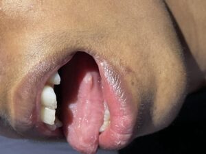
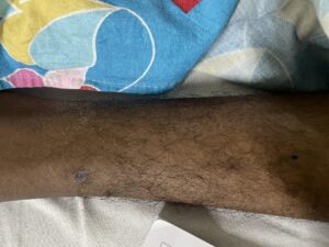
There was a 1 x 1 cm oral aphthous ulcer on the lateral part of the tongue with a well-defined border, and a genital ulcer over the scrotum measuring 1 x 1 cm with a whitish-yellow base .Erythematous plaques were noted over bilateral limbs. Systemic examination was within normal limits.
Labs
Normocytic normochromic anemia (Hb-10.8), with high CRP (79) and ESR (103).


Differential Diagnosis
• Polyarticular Juvenile Idiopathic Arthritis (JIA)
• Inflammatory Bowel Disease
• Behcet’s Disease
• Systemic Lupus Erythematosus (SLE)
• Reiter’s Syndrome
• Abdominal Tuberculosis
• Herpes Simplex Virus (HSV)
In view of clinical features, examination findings, and lab parameters, Behcet’s disease with inflammatory bowel disease was suspected.
An ultrasound of the abdomen showed mildly edematous bowel with discrete enlarged mesenteric lymph nodes, likely indicating colitis. Endoscopy was normal, but colonoscopy revealed a perianal fleshy skin tag and marked ulceration with granulation tissue in the colon. Cryptitis and crypt abscesses were noted. Fecal calprotectin levels were high (1962). HLA typing for Behcet’s was sent, and reports are awaited. Echocardiogram (Echo), preoperative serology, and Mantoux test were normal.
Based on revised international criteria for Behcet’s disease, the diagnosis was made considering recurrent oral and genital ulcers, acneiform lesions, and episodic inflammatory arthritis. Ophthalmologic evaluation was normal. The patient was initiated on Colchicine (Tab Goutinil 500 micrograms BD), 5-ASA, and PPI (Pantop 40 mg). Proper dietary nutrition was advised along with regular follow-up.
DISCUSSION:
Behcet’s disease (BD) is a systemic vasculitis affecting vessels of all sizes, leading to a diverse array of symptoms including recurrent oral, genital, and intestinal ulcers, and skin lesions.
The “belt” of Behcet’s disease is commonly referred to as the “Silk Road” region, encompassing countries like Turkey, Iran, and Japan, although it can occur globally. Clinical diagnosis relies on recognizing recurrent inflammatory attacks and the absence of specific biomarkers or genetic tests.
Oral and genital ulcers, hallmark features of Behcet’s disease, present as recurrent, painful sores significantly impacting quality of life. Oral ulcers typically heal without scarring, while genital ulcers may heal with scarring
Cardiac manifestations can include intracardiac thrombosis, coronary arteritis, and myocardial infarction, while vascular complications often involve arterial or venous thrombosis.
Treatment is tailored to disease activity and severity, using immunomodulatory agents, immunosuppressives, and biologics.
Ocular involvement is common, presenting as anterior or posterior uveitis or retinal vasculitis, while neurological symptoms can include brainstem or pyramidal tract signs.. Treatment strategies include topical corticosteroids, antiseptics, systemic treatments like colchicine, oral corticosteroids, immunosuppressive agents, and biologics. Pain management, good oral hygiene, dietary adjustments, and regular follow-ups are crucial for comprehensive care.
