
Dr.Venkatesh Kumar M, Dr. Praveena N Bhaskaran, Dr.Preethi, DrCJayakumar AIMS,Kochi.
Twelve year old male child, developmentally normal and immunised who is a known case of Supracardiac TAPVC post surgical repair presented with swelling noted in the right posterior triangle of neck of one month duration , initially the size of a coin (0.5 x 0.5 cm), soft later progressed to a size of 4 x 4 cm, hard in consistency.
He also complains of fever associated with night sweats of 3 days duration after 1 week of onset of swelling.
History of dry cough of two weeks duration .He had a dental extraction 1 month prior to onset of swelling.
History of travel to Hep A+ endemic area,1 month back.
He also has history of intake of unpasteurized milk.
USG neck done outside showed – Coarse thyroid echotexture with Lesion in Right posterior triangle- ? Peripheral nerve sheath tumour- Schwannoma.
He was born preterm (35 weeks) via LSCS (Indication: PID- Fallopian tube infection) with birth weight of 2.45 kg.
At 1.5 years, with the complaints of breathing difficulty, cardiac evaluation with ECHO showed SupracardiacTAPVC and he was operated for the same at 2years.
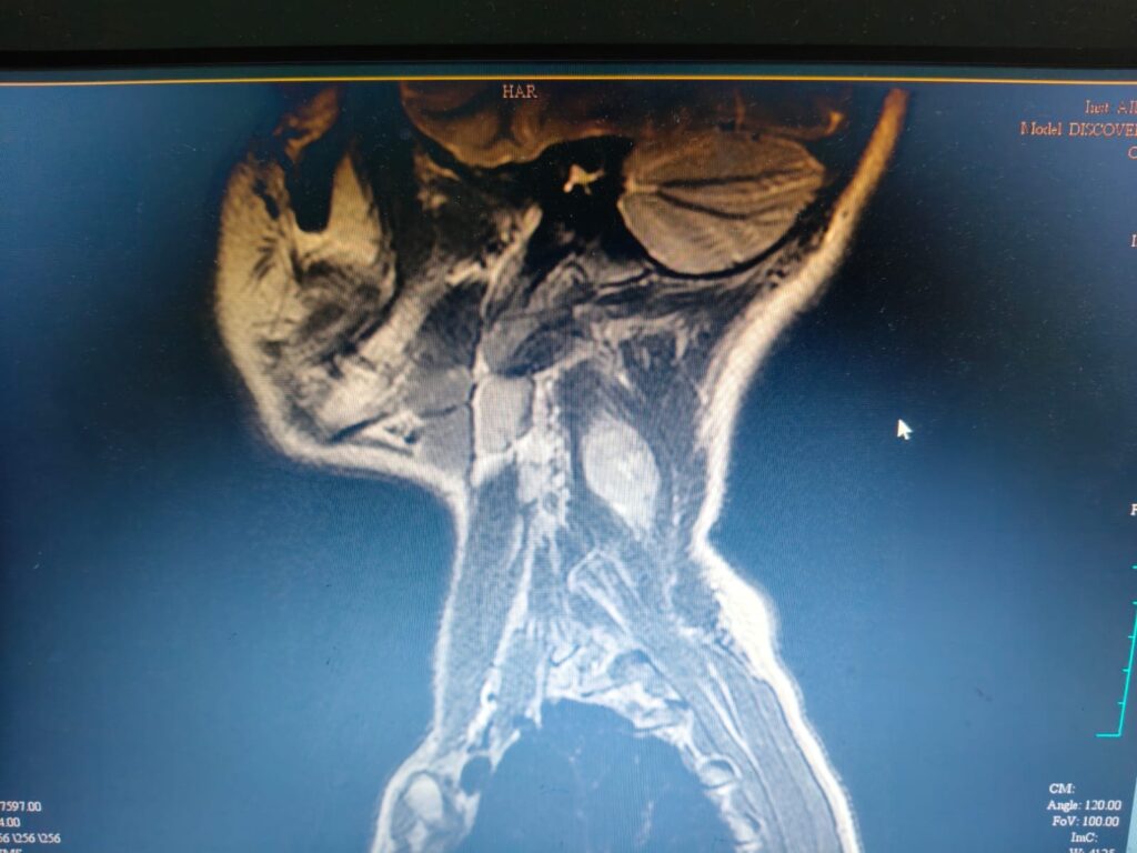
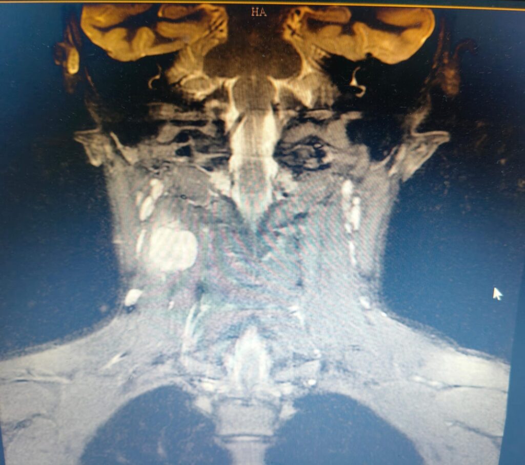
Clinical showed swelling in the Right posterior triangle of neck measuring 4x 4 cm, non tender, firm to hard in consistency, non mobile.
Auxology revealed normal growth parameters.
Systemic examination was unremarkable.
Differentials considered at this point are:
1. Infectious mononucleosis
2. Tuberculosis
3. Strep pharyngitis and secondary lymphadenitis
4. Cat-scratch disease
5. Toxoplasmosis
6 .Benign smooth muscle neoplasms
Investigations:
Hemogram: TC-7.78 ku/ml, N/L: 47.3/ 34.3%, E- 11.1%, Hb-14.4 gm/dl, Plt- 5.43 lakh.
Peripheral smear: Mild Eosinophilia with thrombocytosis.
LFT, RFT: Normal.
HbsAg, Anti-HCV, HIV, Toxoplasma IgM : Negative
EBV IgM: Equivocal.
LDH: 194 U/L.
USG Neck:
A heterogeneous appearing solid lesion in the right posterior triangle,with no features of active inflammation – ? nodal mass.
Suggested HPE correlation. Thyroid gland shows features of thyroiditis.
USG Abdomen: Borderline splenomegaly.
Quantiferon TB gold: Negative.
Biopsy from cervical lymph node and deep cervical mass taken which showed
Cervical lymph node: Paracortical proliferation
Deep cervical mass: Benign spindle cell neoplasm, morphology favouring Proliferative fasciitis
Bacterial and AFB Culture from the lymph node tissue: No growth.
Impression: Spindle cell neoplasm.

Discussion:
Spindle cell and fibrohistiocytic soft tissue tumours are a diverse group of neoplasms that are commonly seen in childhood. Fibroblastic and myofibroblastic tumours are characterized the proliferation of fibroblasts or myofibroblasts, and they exhibit a wide range of histological appearances, from benign to malignant, and can occur in various anatomical locations. Nerve sheath tumours are derived from the Schwann cells of the peripheral nerves and are often associated with neurofibromatosis. Synovial sarcoma, a high-grade malignant tumour, is characterized specific chromosomal translocationsresulting in the SS18-SSX fusion gene. NTRK-rearranged tumours, a recently recognized entity, are characterized gene fusions involving the neurotrophic tyrosine receptor kinase (NTRK) genes, leading to overexpression of the TRK protein. Fibrohistiocytic tumours of childhood, including fibrous histiocytoma and plexiform fibrohistiocytic tumour, show varying degrees of fibroblastic and histiocytic differentiation. They exhibit a wide range of clinical behaviour, from benign to locally aggressive. Understanding the histopathological features of these tumours is important to guide appropriate management.
Carry home message: The diagnosis of cutaneous and subcutaneous spindle cell neoplasms in children is often challenging and has potential therapeutic and prognostic implications.