
Dr Adeena , Dr Naveen, Dr Pramod [Pediatric surgery] , Dr Jayakumar
AIMS Kochi
Three month old boy, 2nd child of NCM, with antenatal history of polyhydraminos and Gestational diabetes on diet control from 5 month of gestation,born preterm 36+1/7 via Emergency LSCS weighing 3kg and had secondary apnoea requiring Positive pressure ventilation followed NICU stay of 27 days ,initially on CPAP and was slowly weaned to room air day 27 of life.
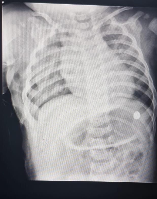
Chest xray soon after birth showed right middle and lower lung zone homogenous opacity with air bronchogram and was managed as congenital pneumonia with Parenteral antibiotics.However as respiratory distress persisted ultrasound of lung showed collapse consolidation, and CT chest showed congenital overinflation/lobar emphysema of right middle lobe.
Post discharge ba was gaining weight but had subcostal retraction which was more prominent after feeds or crying.
The ba was brought to AIMS for further manangement.
O/e: Fronatanel at level.
Wt 6.6 kg at 3months
Air entry decreased on right infrascapular region,subcostal chest retractions while feeding.
Ba underwent lung perfusion scan that showed significantly reduced perfusion to right middle lobe with percentage Tc-99m macro aggregated albumin ( MAA )uptake in segments of RML of 8%.He underwent right middle lobe lobectomy.
Operative finding showed grossly distended RML with good expansion of upper and lower lobe post lobectomy.Histopathological examination of lung tissue showed distended alveloar sac with hemorrhage,no evidence of granuloma /malignancy .
This was consistent with diagnosis of congenital lobar emphysema.
He had an uneventful post operative period and was discharged with stable vitals.
Congenital lobar emphysema (CLE) can present in the neonatal period with respiratory distress, tachypnea, cyanosis, or may be asymptomatic. In infants, it may manifest as wheezing, recurrent respiratory infections, or failure to thrive, whereas in older children, it is often detected incidentally on chest X-rays. Antenatal findings on ultrasound may show a hyperlucent area in the lung with a mediastinal shift. The pathogenesis involves either mechanical factors, such as bronchial cartilage defects or intrinsic airway obstruction, or functional factors like alveolar overexpansion due to extrinsic bronchial compression or anomalous pulmonary vascularity. Diagnosis is based on clinical examination revealing respiratory distress and diminished breath sounds over the affected lobe, supported imaging studies. Chest X-rays typically show a hyperinflated lobe, mediastinal shift, and atelectasis of adjacent lobes, while CT scans offer a detailed assessment of lung anatomy and the extent of involvement. Evaluation includes imaging, lung perfusion ventilation scan and pulmonary function tests(in older children). Complications of CLE can include respiratory distress syndrome from lung tissue compression, recurrent infections due to impaired ventilation, pulmonary hypertension secondary to chronic lung disease, and cardiac compression from mediastinal shift. Management usually involves surgical resection (lobectomy) of the affected lobe in symptomatic cases, though conservative management with close monitoring may be appropriate for asymptomatic patients.
Congenital lobar emphysema (CLE) can present in the neonatal period with respiratory distress, tachypnea, cyanosis, or may be asymptomatic. In infants, it may manifest as wheezing, recurrent respiratory infections, or failure to thrive, whereas in older children, it is often detected incidentally on chest X-rays. Antenatal findings on ultrasound may show a hyperlucent area in the lung with a mediastinal shift. The pathogenesis involves either mechanical factors, such as bronchial cartilage defects or intrinsic airway obstruction, or functional factors like alveolar overexpansion due to extrinsic bronchial compression or anomalous pulmonary vascularity. Diagnosis is based on clinical examination revealing respiratory distress and diminished breath sounds over the affected lobe, supported imaging studies. Chest X-rays typically show a hyperinflated lobe, mediastinal shift, and atelectasis of adjacent lobes, while CT scans offer a detailed assessment of lung anatomy and the extent of involvement. Evaluation includes imaging, lung perfusion ventilation scan and pulmonary function tests(in older children). Complications of CLE can include respiratory distress syndrome from lung tissue compression, recurrent infections due to impaired ventilation, pulmonary hypertension secondary to chronic lung disease, and cardiac compression from mediastinal shift. Management usually involves surgical resection (lobectomy) of the affected lobe in symptomatic cases, though conservative management with close monitoring may be appropriate for asymptomatic patients.