Dr Anakha V Ajay
Dr Jayasree (Neo)DrNaveen Viswa nath( Paed Surgery )
Dr C Jayakumar
AIMS Kochi
Two day old ba born with moral perinatal history did not pass meconium 24 hours of life ba was admitted to NICU for observation.
Ba was also noted to have abdominal distension.
Rectal stimulation with glycerine enema was given and ba passed meconium.
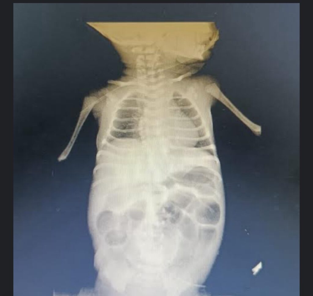
Xray and USG abdomen was suggestive of lower intestinal obstruction.
Ba was also noted to have bilious vomiting for which was kept nil orally and started on potential fluids . Ba was referred here for further management.
On examination, ba was alert with stable vitals. No obvious external anomalies noted. Abdomen examination revealed distended abdomen .Other systemic examination were within normal limits.
Differentials considered were:
1. Hirschprung’s disease
2. Ileojejunal atresia
3. Malrotation
In NICU Ba was hemodynamically stable without any inotropic support, on room air. Ba was initially started on partial parenteral nutrition. NG aspirate were replaced .Barium enema showed imaging features representing long segment Hirschsprung’s disease with transition at splenic flexure. Ba was taken up for surgery and intraoperatively dilated and narrowed small bowel seen:
Bowel resection done with end to end anastomosis was done. Excision biopsy sent from full thickness dilated and narrowed small bowel showed unremarkable mucosa. Nerve bundles and ganglion cells are seen in submucosa. Ganglion cells are also noted in the myenteric plexus. Features keeping with small bowel atresia. Feeds were started from post operative day 6 and was gradually increased as tolerated. In view of sepsis, blood culture was taken and ba was continued on IV Meropenem and Amikacin. Sepsis screen was negative. Antibiotics were stopped after 10 days. Prophylactic antifungals were given in view of central line in situ . Ba was on full feeds day 14 of life. Feed tolerated well the ba. Ba was active, with normal tone. Neurosonogram done was suggestive of Small resolving grade one intraventricular haemorrhage in the left with cystic change.Ba has been enrolled into the early intervention program. Presently ba is on direct breast feeds with multivitamin supplements. Satisfactory weight gain noted and transferred to mother’s side on day 17 of life.At discharge ba is active, feeding well and advised to be under regular follow up.

Ileal and jejunal atresia are usually described together as jejuno ileal atresia (JIA). JIA is a common cause of intestinal obstruction in neonates. It is seen in 1 in 5000 to 1 in 14000 live births. Intestinal atresia can occur in any location on the small bowel as a solitary or even multiple lesions. The cause of jejunoileal atresia has been attributed to an intrauterine vascular accident involving branches of mesenteric vessels in the midgut.
Grosfeld classification:
Type I atresia -internal membrane with serosa continuity and no mesenteric defect;
Type II -involves a proximal and distal blind pouch connected a fibrous cord with serosal discontinuity
Type IIIa- serosa discontinuity with a V-shaped mesenteric defect only.
Type IIIb – apple peel deformity which described proximal jejunal atresia and a short ileal segment coiled around the ileocolic artery.
Type IV-multiple atresias .
Intestinal atresia may be suspected in-utero with suspicious prenatal ultrasound findings, and in a neonate, obstructive symptoms such as abdominal distension, bilious emesis are usually the presenting symptoms. Postnatally, JIA presents with signs and symptoms of intestinal obstruction such as abdominal distension, emesis, and in some cases, delayed passage of meconium. Radiographic examination of the abdomen with plain abdominal X-ray using swallowed air as a contrast is a useful diagnostic tool. For proximal JIA, there are presences of a few dilated proximal bowel with no distal gas.The most common technique is resection of proximal dilated and atretic bowel with primary end-to-end anastomosis with or without tapering enteroplasty of the proximal bowel.
Bowel loss is more common in type IIIb and type IV atresia. The prognosis of JIA depends on the presence of short bowel syndrome (SBS) with an intestinal length of less than 25cm, requiring long-term parenteral nutrition.
Take home message-Multidisciplinary care improves outcomes and standardized protocols enhance diagnosis and treatment
