Dr. Venkat Kumar Raju, Dr. Vaishakh Anand, Dr.Jayakumar
Fifteen yr old with normal antenatal and developmental background who is a known case of cerebellar pilocytic astrocytoma (WHO GRADE 1), S/P Sub-occipital craniotomy and near total excision of the lesion presented with the complaints of paroxysmal event in form of up rolling of eyeballs and tonic clonic movements of all 4 limbs , which were managed with levteracetam .
One week later parents noticed tremors in tongue ,tremors were also present during awake and sleep, got worsened during eating and noticed to have drooling of saliva.
Past history – at 14 years due to head ache and early morning vomiting MRI was done , and was operated within a month
On Examination
CNS – HMF- conscious and coherent ,
Cranial nerve showed left lateral rectus palsy , bilateral ptosis and bilateral facial nerve palsy
Motor- bulk normal, hypotonia , power 5/5, DTR normal
Has nystagmus, slurred speech, tandem walking impaired and ataxic gait , tongue tremors were seen
EEG showed non specific disturbances of electrical function over bilateral frontal region
Paraneoplastic and autoimmune encephalitis panel was negative.
As there is temporal correlation between initiation of tremors and initiation of levipil. It was tapered and switched to oxcarbazepine
The overall clinical picture is more suggestive of tongue and postal tremors – Giullian Mollarettriangle lesion was suspected
the child was started on Gabapentin

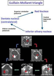
Giulian Mollaret Triangle
The triangle of Guillain and Mollaret, also known as the dentatorubro-olivary pathway, has three corners 1:
- red nucleus
- inferior olivary nucleus
- contralateral dentate nucleus
Rubro-olivary fibers descend from the parvocellular division of each red nucleus along the central tegmental tracts to reach the capsule (amiculum) of the ipsilateral inferior olivary nucleus (ION). From the ION, olivocerebellar fibers cross the contralateral inferior cerebellar peduncle to reach cerebellar cortex, then pass from cerebellar cortex to the contralateral dentate nucleus. Dentatorubral fibers then ascend via the contralateral superior cerebellar peduncle, decussate in the midbrain, and return to the original red nucleus.
No direct connecting tract is present between the inferior olivary nucleus and contralateral dentate nucleus.
History and etymology
The pathway was described in 1931 the French neurologists Georges Charles Guillain (1876-1961) and Pierre Mollaret (1898-1987).They are also known respectively for defining what is now known as Guillain-Barré syndrome and Mollaret meningitis.
Related pathology - hypertrophic olivary degeneration, manifest as palatal myoclonus
o contralateral to lesions of the superior cerebellar peduncle
o ipsilateral to lesions of the central tegmental tract - cerebellar atrophy
o contralateral to lesions of the olivocerebellar fibers - Holmes tremor (double lesions in both the dentatorubral-olivary system and dopaminergic nigrostriatal system)
- Treatment for lesions in the GMT is usually symptomatic. Medications like gabapentin, memantine, and trihexyphenidyl can help manage symptoms like palatal tremor, eye oscillations, and pupil oscillations.
- Prognosis Most patients with symptomatic hypertrophic olivary degeneration (HOD) see their symptoms improve over time. Some patients can relieve their symptoms themselves after 3 to 4 years
